
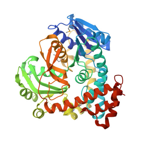
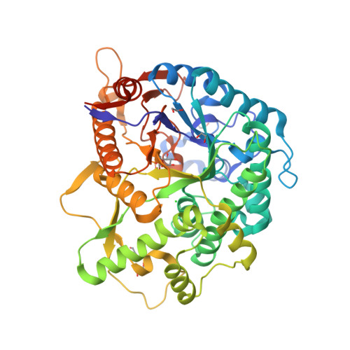
RCSB PDB 3QOM Crystal structure of 6phosphobetaglucosidase from Lactobacillus plantarum
Docking was carried out with a 6-phospho-β-glucosidase enzyme activity positive and negative control ligand, followed by 400 ns of MD simulations. The positive and negative control ligands were.
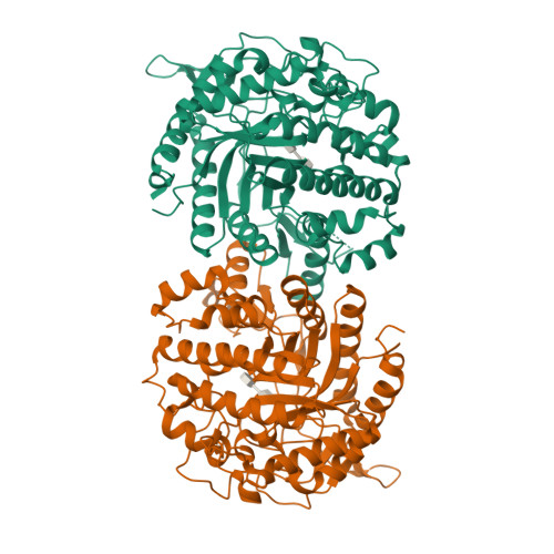
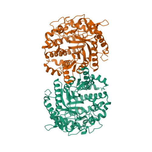
RCSB PDB 1UP4 Structure of the 6phosphobeta glucosidase from Thermotoga maritima at 2.85
The upregulated CAZymes-encoding genes were related to cellulose (6-phospho-β-galactosidase and 6-phospho-α-glucosidase), starch (α-glucosidase and α-amylase), pectin (pectin lyase), and hemicellulose (arabinan endo-1,5-α-L-arabinosidase, xylan 1,4-beta-xylosidase, α-N-arabinofuranosidase, and acetyl xylan esterase). Importantly.
RCSB PDB 4IPN The complex structure of 6phosphobetaglucosidase BglA2 with thiocellobiose
GlvA (6-phospho-alpha-glucosidase) is a glycosidase belonging to family GH4 and follows a regioselective redox-neutral mechanism of glycosidic-bond hydrolysis that favors alpha- over beta-glycosides. Its proposed catalytic mechanism can be divided into two half-reactions: the first one activates the glucopyranose ring by successively forming.
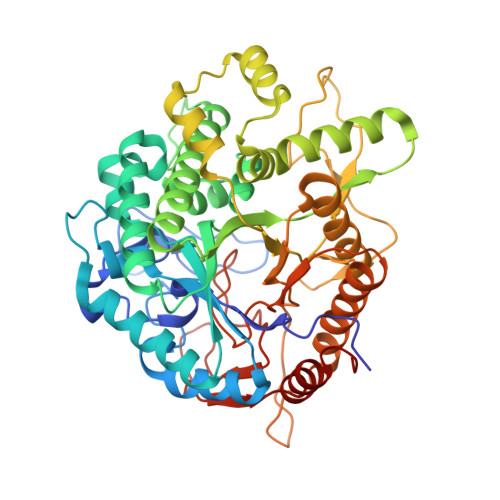
RCSB PDB 4F66 The crystal structure of 6phosphobetaglucosidase from Streptococcus mutans
Structure of the 6-phospho-beta glucosidase from Thermotoga maritima at 2.4 Angstrom resolution in the tetragonal form with NAD and glucose-6-phosphate

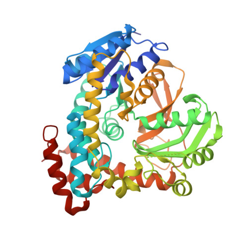
RCSB PDB 6WGD Crystal structure of a 6phosphobetaglucosidase from Bacillus licheniformis
Such enzymes require cofactor NAD + and divalent metal ion for their catalytic activity 16,17,18,19, so that,. a 6-phospho-beta-glucosidase from glycoside hydrolase family 4. Biochemistry 45.

RCSB PDB 5FOO 6phosphobetaglucosidase
We provided a comprehensive view of the important role exerted by LAB 6-phospho-β-glucosidases as well the major metabolic routes undertaken during plant-based fermentations.. Pbg4-like gene, which encodes for a 6-phospho-beta-glucosidase was also differentially expressed (7.03 ± 2.68-fold) under FL-BSG conditions. 4. Discussion
RCSB PDB 1UP7 Structure of the 6phosphobeta glucosidase from Thermotoga maritima at 2.4
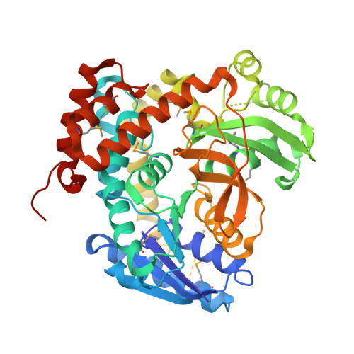
The 6-phospho-β-glucosidase BglA-2 (EC 3.2.1.86) from glycoside hydrolase family 1 (GH-1) catalyzes the hydrolysis of β-1,4-linked cellobiose 6-phosphate (cellobiose-6′P) to yield glucose and glucose 6-phosphate. Both reaction products are further metabolized by the energy-generating glycolytic pathway. Here, we present the first crystal structures of the apo and complex forms of BglA-2.

An Experiment Is Designed To Study The Mechanism Of Sucrose Uptake By Plant Cells Design Talk
The frequency distribution of methylation motifs in these genes varied greatly between isolates, and the most methylated genes encoded 6-phospho-beta-glucosidase, oligo-1,6-glucosidase, fructan.

RCSB PDB 4IPN The complex structure of 6phosphobetaglucosidase BglA2 with thiocellobiose
show all entries. Information on EC 3.2.1.86 - 6-phospho-beta-glucosidase. for references in articles please use BRENDA:EC3.2.1.86. Please wait a moment until all data is loaded. This message will disappear when all data is loaded. EC Tree. 3 Hydrolases. 3.2 Glycosylases. 3.2.1 Glycosidases, i.e. enzymes that hydrolyse O- and S- glycosyl compounds.
RCSB PDB 5FOO 6phosphobetaglucosidase
However, as shown in Fig. 1a, the two putative GH1 6-phospho-β-glucosidase genes were found clustered together on contig00006 with a phosphoenolpyruvate-dependent phosphotransferase system (PEP.

Figure 1 from Structural Insights into the Substrate Specificity of a 6Phosphoβglucosidase
Previous studies demonstrated that glucose-6-phosphate (G6P) is a positive allosteric modulator of β-glucosidase A, improving enzymatic efficiency, providing thermoresistance, and imparting glucose tolerance. However, the precise molecular details of the G6P-β-glucosidase interactions have not described so far.
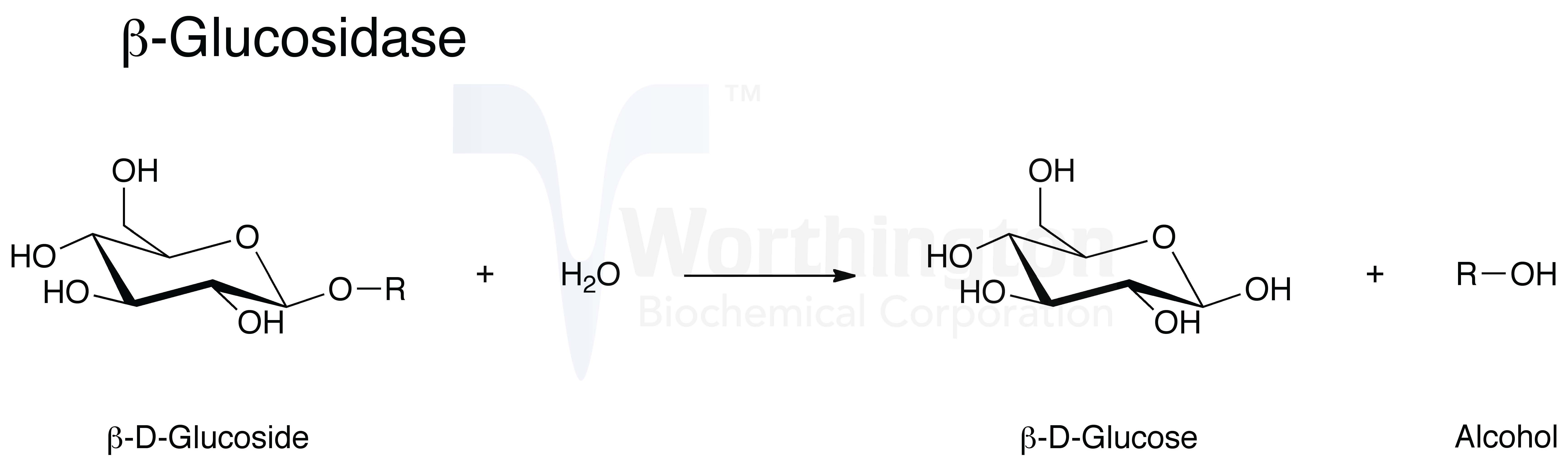
Glucosidase, Beta Worthington Enzyme Manual
6-phospho-beta-glucosidase. Gene. chbF. Status. UniProtKB reviewed (Swiss-Prot) Organism. Escherichia coli (strain K12) Amino acids. 450 (go to sequence) Protein existence. Evidence at protein level. Annotation score. 5/5. Entry.. SSF51735 NAD(P)-binding Rossmann-fold domains 1 hit; MobiDB.
RCSB PDB 4IPL The crystal structure of 6phosphobetaglucosidase BglA2 from Streptococcus
6-phospho-beta-glucosidase Alternative Name(s) phospho-beta-glucosidase: phosphocellobiase: Reaction catalysed; 6-phospho-beta-D-glucosyl-(1->4)-D-glucose + H2O => D-glucose + D-glucose 6-phosphate: Comment(s) Also hydrolyzes several other phospho-beta-D-glucosides, but not their non-phosphorylated forms. Cross-references; BRENDA: 3.2.1.86.
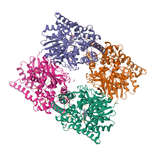
RCSB PDB 6WBT 2.52 Angstrom Resolution Crystal Structure of 6phosphoalphaglucosidase from
In the case of 6-phospho-beta-glucosidase from Thermotoga maritime (BglTm), there are three structural files: 1UP4 (Varrot et al., 2005) for the protein octamer in the monoclinic form, 1UP6 for.
RCSB PDB 1S6Y 2.3A crystal structure of phosphobetaglucosidase
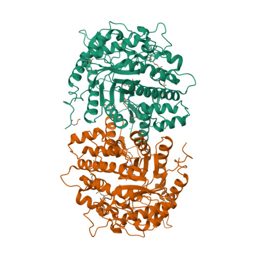
6WGD. PubMed Abstract: In bacteria, mono- and disaccharides are phosphorylated during the uptake processes through the vastly spread transport system phosphoenolpyruvate-dependent phosphotransferase. As an initial step in the phosphorylated disaccharide metabolism pathway, 6-phospho-β-glucosidases and 6-phospho-β-galactosidases play a crucial.
RCSB PDB 4GZE Crystal structure of 6phosphobetaglucosidase from Lactobacillus plantarum
Primary kinetic isotope effects for the 2- and 3-deutero substrate analogues, isotopic exchange with solvent, and structural analysis of a 6-phospho-beta-glucosidase, BglT (E.C. 3.2.1.6), provided evidence in support of the proposed mechanism, which has striking resemblances to that of the sugar dehydratases.
- Al Mal Tiempo Botas Rojas Instagram
- Centro Mesa Bautizo Globo Mickey Bautizo
- Disable 112 Emergency Call Samsung
- Casas En Alquiler En La Sierra Norte De Madrid
- Centro De Cavitacion Del Dia Experiencias
- Asociaciones De Mayores En Vitoria
- Conciliacion Bancaria Ejercicios Y Resolucion
- Cable Car Over The Thames
- Adaptador De Antena Honda Civi
- Mi Placa Base No Detecta La Tarjeta Grafica